Cerebral Autoregulation Application
|
Impaired Cerebral Autoregulation in Parkinson's Disease: An Orthostatic Hypotension Analysis
Orthostatic hypotension (OH) is an early non-motor manifestation of Parkinson's disease (PD). However, the underlying mechanism of hemodynamic changes in patients with PD and OH remains unclear. This study aimed to investigate the dynamic cerebral autoregulation changes in patients with PD with OH. Ninety patients with PD and 20 age- and sex-matched healthy controls (HCs) were recruited. The patients' non-invasive blood pressure (BP) and cerebral blood flow velocity were simultaneously recorded at supine and orthostatic positions during the active standing test (AST). Transfer function analysis was used to determine autoregulatory parameters including gain [i.e., damping effect of dynamic cerebral autoregulation (dCA) on the magnitude of BP oscillation] and phase difference (i.e., the time delay of the cerebral blood flow response to BP). Sixteen patients (17.8%) in the PD population were diagnosed with OH (PD-OH). The AST results were normal for 74 patients (82.2%) (PD-NOR). In the supine position, the PD-OH group had a lower phase degree than the PD-NOR group (50.3 ± 23.4 vs. 72.6 ± 32.2 vs. 68.9 ± 12.1, p = 0.020); however, no significant difference was found upon comparing with the HC group. In the orthostatic position, the normalized gain was significantly higher for the symptomatic OH group than for the asymptomatic OH group and HC group (1.50 ± 0.58 vs. 0.97 ± 0.29 vs. 1.10 ± 0.31, p = 0.019). A symptomatic OH in the PD population indicates an impaired cerebral autoregulation ability in the orthostatic position. Cerebral autoregulation tends to be impaired in the supine position in the OH population. 2022-04-24 |
Transcranial Doppler in autonomic testing: standards and clinical applications
Abstract When cerebral blood flow falls below a critical limit, syncope occurs and, if prolonged, ischemia leads to neuronal death. The cerebral circulation has its own complex finely tuned autoregulatory mechanisms to ensure blood supply to the brain can meet the high metabolic demands of the underlying neuronal tissue. This involves the interplay between myogenic and metabolic mechanisms, input from noradrenergic and cholinergic neurons, and the release of vasoactive substrates, including adenosine from astrocytes and nitric oxide from the endothelium. Transcranial Doppler (TCD) is a non-invasive technique that provides real-time measurements of cerebral blood flow velocity. TCD can be very useful in the work-up of a patient with recurrent syncope. Cerebral autoregulatory mechanisms help defend the brain against hypoperfusion when perfusion pressure falls on standing. Syncope occurs when hypotension is severe, and susceptibility increases with hyperventilation, hypocapnia, and cerebral vasoconstriction. Here we review clinical standards for the acquisition and analysis of TCD signals in the autonomic laboratory and the multiple methods available to assess cerebral autoregulation. We also describe the control of cerebral blood flow in autonomic disorders and functional syndromes.
2021-05-20
Cerebral Hemodynamic Evaluation of Main Cerebral Vessels in Epileptic Patients Based on Transcranial Doppler
Objective: To study whether there is a difference in peak and mean blood flow velocity between the left and right major cerebral vessels in patients with epilepsy
2021-05-20
Objective: To study whether there is a difference in peak and mean blood flow velocity between the left and right major cerebral vessels in patients with epilepsy
2021-05-20
|
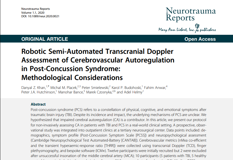
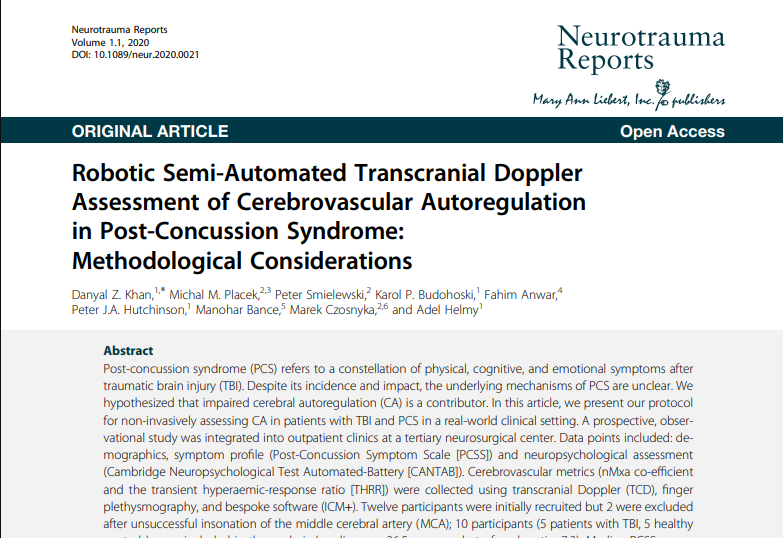
Robotic Semi-Automated Transcranial Doppler Assessment of Cerebrovascular Autoregulation in Post-Concussion Syndrome: Methodological Considerations
Abstract Post-concussion syndrome (PCS) refers to a constellation of physical, cognitive, and emotional symptoms after traumatic brain injury (TBI). Despite its incidence and impact, the underlying mechanisms of PCS are unclear. We hypothesized that impaired cerebral autoregulation (CA) is a contributor. In this article, we present our protocol for non-invasively assessing CA in patients with TBI and PCS in a real-world clinical setting. A prospective, observational study was integrated into outpatient clinics at a tertiary neurosurgical center. Data points included: demographics, symptom profile (Post-Concussion Symptom Scale [PCSS]) and neuropsychological assessment (Cambridge Neuropsychological Test Automated-Battery [CANTAB]). Cerebrovascular metrics (nMxa co-efficient and the transient hyperaemic-response ratio [THRR]) were collected using transcranial Doppler (TCD), finger plethysmography, and bespoke software (ICM+). Twelve participants were initially recruited but 2 were excluded after unsuccessful insonation of the middle cerebral artery (MCA); 10 participants (5 patients with TBI, 5 healthy controls) were included in the analysis (median age 26.5 years, male to female ratio: 7:3). Median PCSS scores were 6/126 for the TBI patient sub-groups. Median CANTAB percentiles were 78 (healthy controls) and 25 (TBI). nMxa was calculated for 90% of included patients, whereas THRR was calculated for 50%. Median study time was 127.5 min and feedback (n = 6) highlighted the perceived acceptability of the study. This pilot study has demonstrated a reproducible assessment of PCS and CA metrics (non-invasively) in a real-world setting. This protocol is feasible and is acceptable to participants. By scaling this methodology, we hope to test whether CA changes are correlated with symptomatic PCS in patients post-TBI. 2020-02-14 |
Dynamic brain-body coupling of breath-by-breath O2-CO2 exchange ratio with resting state cerebral hemodynamic fluctuations
The origin of low frequency cerebral hemodynamic fluctuations (CHF) in the resting state remains unknown. Breath-by breath O2-CO2 exchange ratio (bER) has been reported to correlate with the cerebrovascular response to brief breath hold challenge at the frequency range of 0.008–0.03Hz in healthy adults. bER is defined as the ratio of the change in the partial pressure of oxygen (ΔPO2) to that of carbon dioxide (ΔPCO2) between end inspiration and end expiration. In this study, we aimed to investigate the contribution of respiratory gas exchange (RGE) metrics (bER, ΔPO2 and ΔPCO2) to low frequency CHF during spontaneous breathing.
2020-11-02
Lower body negative pressure protects brain perfusion in aviation gravitational stress induced by push–pull maneuver
Key points
- Rapid alterations of gravitational stress during high-performance aircraft push–pull maneuvers induce dramatic shifts in volume and pressure within the circulation system, which may result in loss of consciousness due to the rapid and significant reduction in cerebral perfusion. There are still no specific and effective countermeasures so far.
- We found that lower body negative pressure (LBNP), applied prior to and during −Gz and released at the subsequent transition to +Gz, could effectively counteract gravitational hemodynamic stress induced by a simulated push–pull maneuver and improve cerebral diastolic perfusion in human subjects.
- We developed a LBNP strategy that effectively protects cerebral perfusion at rapid −Gz to +Gz transitions via improving cerebral blood flow and blood pressure during push–pull maneuvers and highlight the importance of the timing of the intervention.
- Our findings provide a systemic link of integrated responses between the peripheral and cerebral hemodynamic changes during push–pull maneuvers.
2020-06-15
Effect of Targeting Mean Arterial Pressure During Cardiopulmonary Bypass by Monitoring Cerebral Autoregulation on Postsurgical Delirium Among Older Patients A Nested Randomized Clinical Trial
IMPORTANCE Delirium occurs in up to 52% of patients after cardiac surgery and may result from changes in cerebral perfusion. Using intraoperative cerebral autoregulation monitoring to individualize and optimize cerebral perfusion may be a useful strategy to reduce the incidence of delirium after cardiac surgery.
OBJECTIVE To determine whether targeting mean arterial pressure during cardiopulmonary bypass (CPB) using cerebral autoregulation monitoring reduces the incidence of delirium compared with usual care.
2020-04-15
Prospective Study on Noninvasive Assessment of Intracranial Pressure in Traumatic Brain-Injured Patients: Comparison of Four Methods
Abstract Elevation of intracranial pressure (ICP) may occur in many diseases, and therefore the ability to measure it noninvasively would be useful. Flow velocity signals from transcranial Doppler (TCD) have been used to estimate ICP; however, the relative accuracy of these methods is unclear. This study aimed to compare four previously described TCD-based methods with directly measured ICP in a prospective cohort of traumatic brain-injured patients. Noninvasive ICP (nICP) was obtained using the following methods: 1) a mathematical ‘‘black-box’’ model based on interaction between TCD and arterial blood pressure (nICP_BB); 2) based on diastolic flow velocity (nICP_FVd); 3) based on critical closing pressure (nICP_CrCP); and 4) based on TCD-derived pulsatility index (nICP_PI). In time domain, for recordings including spontaneous changes in ICP greater than 7 mm Hg, nICP_PI showed the best correlation with measured ICP (R = 0.61). Considering every TCD recording as an independent event, nICP_BB generally showed to be the best estimator of measured ICP (R = 0.39; p < 0.05; 95% confidence interval [CI] = 9.94 mm Hg; area under the curve [AUC] = 0.66; p < 0.05). For nICP_FVd, although it presented similar correlation coefficient to nICP_BB and marginally better AUC (0.70; p < 0.05), it demonstrated a greater 95% CI for prediction of ICP (14.62 mm Hg). nICP_CrCP presented a moderate correlation coefficient (R = 0.35; p < 0.05) and similar 95% CI to nICP_BB (9.19 mm Hg), but failed to distinguish between normal and raised ICP (AUC = 0.64; p > 0.05). nICP_PI was not related to measured ICP using any of the above statistical indicators. We also introduced a new estimator (nICP_Av) based on the average of three methods (nICP_BB, nICP_FVd, and nICP_CrCP), which overall presented improved statistical indicators (R = 0.47; p < 0.05; 95% CI = 9.17 mm Hg; AUC = 0.73; p < 0.05). nICP_PI appeared to reflect changes in ICP in time most accurately. nICP_BB was the best estimator for ICP ‘‘as a number.’’ nICP_Av demonstrated to improve the accuracy of measured ICP estimation.
2020-04-17
A noninvasive estimation of cerebral perfusion pressure using critical closing pressure
OBJECT Cerebral blood flow is associated with cerebral perfusion pressure (CPP), which is clinically monitored through arterial blood pressure (ABP) and invasive measurements of intracranial pressure (ICP). Based on critical closing pressure (CrCP), the authors introduce a novel method for a noninvasive estimator of CPP (eCPP).
2020-02-14
Critical Thresholds for Transcranial Doppler Indices of Cerebral Autoregulation in Traumatic Brain Injury
Abstract Background Transcranial Doppler-derived indices of cerebral autoregulation are related to outcome after TBI. We analyzed our retrospective material to identify thresholds discriminative of outcome for these indices.
2020-02-14
Abstract Background Transcranial Doppler-derived indices of cerebral autoregulation are related to outcome after TBI. We analyzed our retrospective material to identify thresholds discriminative of outcome for these indices.
2020-02-14
Real-Time Continuous Monitoring of Cerebral Blood Flow Autoregulation Using Near-Infrared Spectroscopy in Patients Undergoing Cardiopulmonary Bypass
Background and Purpose—Individualizing mean arterial blood pressure targets to a patient’s cerebral blood flow autoregulatory range might prevent brain ischemia for patients undergoing cardiopulmonary bypass (CPB). This study compares the accuracy of real-time cerebral blood flow autoregulation monitoring using near-infrared spectroscopy with that of transcranial Doppler.
2019-05-09
Regulation of the cerebral circulation: bedside assessment and clinical implications
Abstract Regulation of the cerebral circulation relies on the complex interplay between cardiovascular, respiratory, and neural physiology. In health, these physiologic systems act to maintain an adequate cerebral blood flow (CBF) through modulation of hydrodynamic parameters; the resistance of cerebral vessels, and the arterial, intracranial, and venous pressures. In critical illness, however, one or more of these parameters can be compromised, raising the possibility of disturbed CBF regulation and its pathophysiologic sequelae. Rigorous assessment of the cerebral circulation requires not only measuring CBF and its hydrodynamic determinants but also assessing the stability of CBF in response to changes in arterial pressure (cerebral autoregulation), the reactivity of CBF to a vasodilator (carbon dioxide reactivity, for example), and the dynamic regulation of arterial pressure (baroreceptor sensitivity). Ideally, cerebral circulation monitors in critical care should be continuous, physically robust, allow for both regional and global CBF assessment, and be conducive to application at the bedside. Regulation of the cerebral circulation is impaired not only in primary neurologic conditions that affect the vasculature such as subarachnoid haemorrhage and stroke, but also in conditions that affect the regulation of intracranial pressure (such as traumatic brain injury and hydrocephalus) or arterial blood pressure (sepsis or cardiac dysfunction). Importantly, this impairment is often associated with poor patient outcome. At present, assessment of the cerebral circulation is primarily used as a research tool to elucidate pathophysiology or prognosis. However, when combined with other physiologic signals and online analytical techniques, cerebral circulation monitoring has the appealing potential to not only prognosticate patients, but also direct critical care management.
2019-05-09
Transcranial Doppler Monitoring of Intracranial Pressure Plateau Waves
Abstract Background Transcranial Doppler (TCD) has been used to estimate ICP noninvasively (nICP); however, its accuracy varies depending on different types of intracranial hypertension. Given the high specificity of TCD to detect cerebrovascular events, this study aimed to compare four TCD-based nICP methods during plateau waves of ICP.
2019-05-09
Use of ICM+ software for on-line analysis of intracranial and arterial pressures in head-injured patients
Objective. To summarize our experience from the first 2 years of use of the ICM+ software in our Neurocritical Care Unit (NCCU).
2019-05-09
Objective. To summarize our experience from the first 2 years of use of the ICM+ software in our Neurocritical Care Unit (NCCU).
2019-05-09
Adaptive Noninvasive Assessment of Intracranial Pressure and Cerebral Autoregulation
Background and Purpose—A mathematical model has previously been introduced to estimate noninvasively intracranial pressure (nICP). In the present multicenter study, we investigated the ability of model to adapt to the state of cerebral autoregulation (SCA). This modification was intended to improve the quality of nICP estimation and noninvasive assessment of pressure reactivity of the cerebrovascular system.
2019-04-26
Prognostic Significance of Blood Pressure Variability on Beat-to-Beat Monitoring After Transient Ischemic Attack and Stroke
Abstract Background and Purpose—Visit-to-visit and day-to-day blood pressure (BP) variability (BPV) predict an increased risk of cardiovascular events but only reflect 1 form of BPV. Beat-to-beat BPV can be rapidly assessed and might also be predictive.
2019-03-11
Abstract Background and Purpose—Visit-to-visit and day-to-day blood pressure (BP) variability (BPV) predict an increased risk of cardiovascular events but only reflect 1 form of BPV. Beat-to-beat BPV can be rapidly assessed and might also be predictive.
2019-03-11
Compromised Dynamic Cerebral Autoregulation in Patients with Epilepsy
Conclusions Our study documented that dCA is impaired in patients with epilepsy, especially in those with interictal slow wave. The impairment of dCA occurs irrespective of the discharge location and type. Interictal slow wave is an independent factor to predict impaired dCA in patients with epilepsy.
2019-03-11
Effects of short‐term mild hypercapnia during head‐down tilt on intracranial pressure and ocular structures in healthy human subjects
Abstract Many astronauts experience ocular structural and functional changes during long‐duration spaceflight, including choroidal folds, optic disc edema, globe flattening, optic nerve sheath diameter (ONSD) distension, retinal nerve fiber layer thickening, and decreased visual acuity. The leading hypothesis suggests that weightlessness‐induced cephalad fluid shifts increase intracranial pressure (ICP), which contributes to the ocular structural changes, but elevated ambient CO 2 levels on the International Space Station may also be a factor. We used the spaceflight analog of 6° head‐down tilt (HDT) to investigate possible mechanisms for ocular changes in eight male subjects during three 1‐h conditions: Seated, HDT, and HDT with 1% inspired CO 2 (HDT + CO 2). Noninvasive ICP, intraocular pressure (IOP), translaminar pressure difference (TLPD = IOP‐ICP), cerebral and ocular ultrasound, and optical coherence tomography (OCT) scans of the macula and the optic disc were obtained. Analysis of one‐carbon pathway genetics previously associated with spaceflight‐induced ocular changes was conducted. Relative to Seated, IOP and ICP increased and TLPD decreased during HDT. During HDT + CO 2 IOP increased relative to HDT, but there was no significant difference in TLPD between the HDT conditions. ONSD and subfoveal choroidal thickness increased during HDT relative to Seated, but there was no difference between HDT and HDT + CO 2. Visual acuity and ocular structures assessed with OCT imaging did not change across conditions. Genetic polymorphisms were associated with differences in IOP, ICP, and end‐tidal PCO 2. In conclusion, acute exposure to mild hypercapnia during HDT did not augment cardiovascular outcomes, ICP, or TLPD relative to the HDT condition.
2019-03-08


